39 parts of the eye without labels
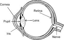
Eye Diagram With Labels and detailed description - BYJUS Iris is the coloured part of the eye and controls the amount of light entering the eye by regulating the size of the pupil. The lens is located just behind the iris. Its function is to focus the light on the retina. The optic nerve transmits electrical signals from the retina to the brain. Pupil is the opening at the centre of the iris. Eye Anatomy: Parts of the Eye and How We See Behind the anterior chamber is the eye's iris (the colored part of the eye) and the dark hole in the middle called the pupil. Muscles in the iris dilate (widen) or constrict (narrow) the pupil to control the amount of light reaching the back of the eye. Directly behind the pupil sits the lens. The lens focuses light toward the back of the eye.
Eye anatomy: A closer look at the parts of the eye Eye anatomy: A closer look at the parts of the eye. By Liz Segre. When surveyed about the five senses — sight, hearing, taste, smell and touch — people consistently report that their eyesight is the mode of perception they value (and fear losing) most. Despite this, many people don't have a good understanding of the anatomy of the eye, how ...
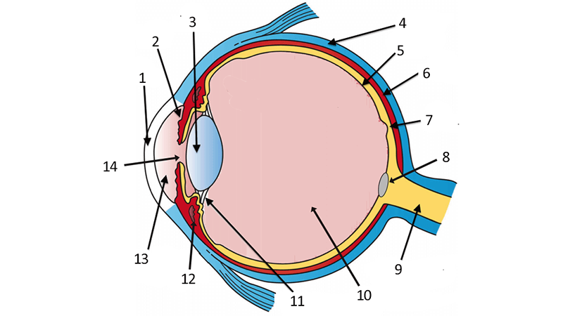
Parts of the eye without labels
› sailboatSailboat Hardware, Sailboat Accessories & Parts | Fisheries ... Sailboat Hardware, Parts & Accessories Fisheries Supply is your premier supplier of sailboat hardware and accessories from top brands like Harken, Ronstan, Lewmar, Schaefer and more. Both online and in our Seattle store, we offer a full range of quality sailboat supplies including winches, rigging parts, blocks, spinnaker poles, shackles ... Label the Parts of the Eye Quiz - PurposeGames.com States Without the Letter 'A' 14p Type-the-Answer. Planets Ordered by Size 9p Image Quiz. ... This is an online quiz called Label the Parts of the Eye. There is a printable worksheet available for download here so you can take the quiz with pen and paper. Your Skills & Rank. Total Points. 0. Eye Diagram Unlabelled - schematron.org 07.12.2018. 1 Comments. on Eye Diagram Unlabelled. Select the correct label for each part of the eye. The image is taken from above the left eye. Click on the Score button to see how you did. Incorrect answers will. 61 high-quality Unlabeled Eye Diagram for free! human eye diagram. YouTube.
Parts of the eye without labels. Eye Diagram - Differentiated Worksheets and EASEL Activities - Pinterest Use this simple eye diagram for primary students as they learn about the human eye. Two differentiated worksheets included: one with a word bank and one without. Words to label: eyebrow, eyelid, eyelashes, pupil, iris, and sclera. Find this Pin and more on Ylli by Kirsi Liuska. Eye Anatomy Diagram. Ear Diagram. Science Student. › custom-labelsCustom Labels For Products - LabelValue We've seen clients do everything from eye-catching point of sale stickers to event promotion stickers to custom water bottle labels for corporate events and more. Our short-run capabilities & SPLASH Variable Design will help your business connect with customers and generate buzz, all without breaking the budget. PDF Eye Anatomy Handout - National Eye Institute of light entering the eye. Lens: The lens is a clear part of the eye behind the iris that helps to focus light, or an image, on the retina. Macula: The macula is the small, sensitive area of the retina that gives central vision. It is located in the center of the retina. Optic nerve: The optic nerve is the largest sensory nerve of the eye. Parts of the Eye & Their Function | Robertson Optical and Optometry The different parts of the eye allow the body to take in light and perceive objects around us in the proper color, detail and depth. This allows people to make more informed decisions about their environment. If a portion of the eye becomes damaged, you may not be able to see effectively, or lose your vision all together.
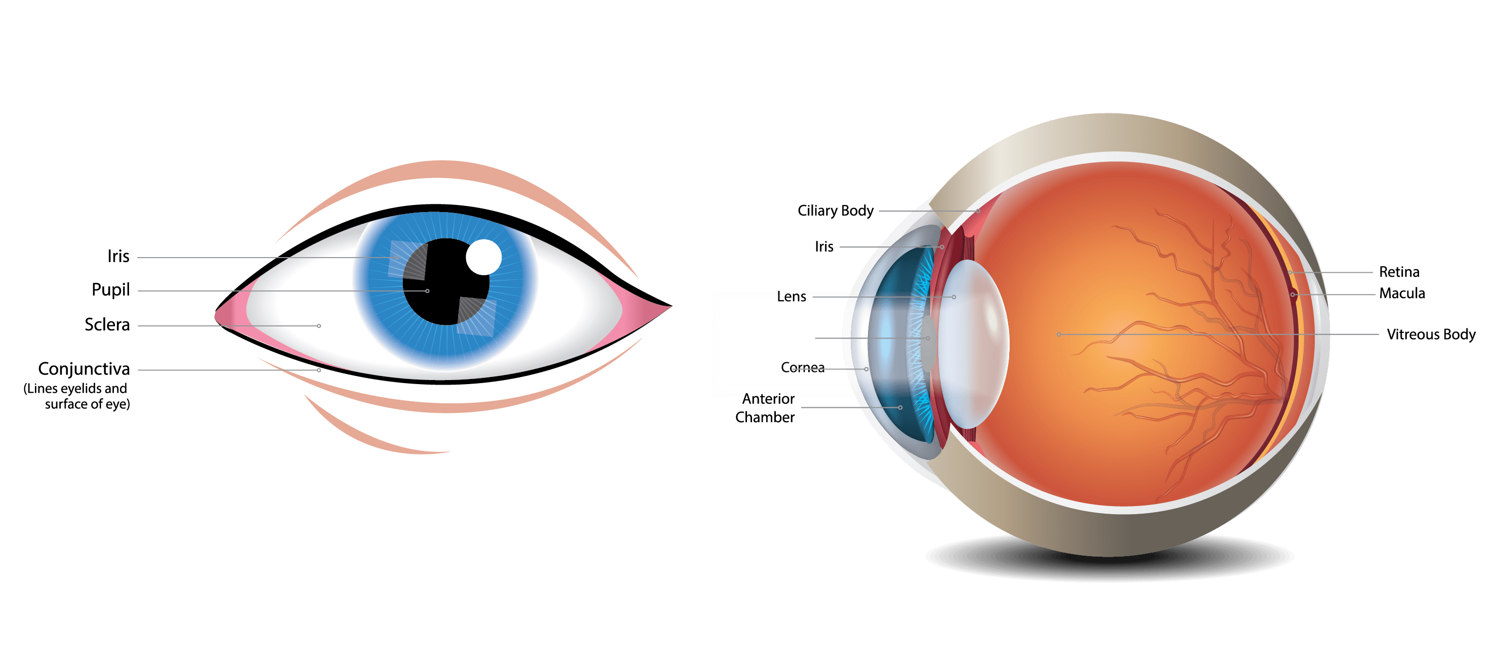
Anatomy of the Eye. Learn about the different parts of the eye. The sclera is a membrane of tendon in the eye, also known as the white of the eye. Rugged and robust, the sclera works to protect the inner, more sensitive parts of the eye like the retina and choroid. It is about 0.03 of an inch thick except for where the four "straight" eye muscles append, where the depth is no more than 0.01 of an inch. Eye Anatomy Detail Picture Image on MedicineNet.com Picture of Eye Anatomy Detail The eye is our organ of sight. The eye has a number of components which include but are not limited to the cornea, iris, pupil, lens, retina, macula, optic nerve, choroid and vitreous. Cornea: clear front window of the eye that transmits and focuses light into the eye. Eye Anatomy: 16 Parts of the Eye & Their Functions - Vision Center The following are parts of the human eyes and their functions: 1. Conjunctiva The conjunctiva is the membrane covering the sclera (white portion of your eye). The conjunctiva also covers the interior of your eyelids. Conjunctivitis, often known as pink eye, occurs when this thin membrane becomes inflamed or swollen. Best Color Label Printer of 2022 – Enterprise Labels The Epson CW-C4000 can help you make good quality labels without breaking the budget. Larger producers needing high volumes of labels may get the entrance color label printer too slow due to the demanding, higher volume manufacturing environments. The Epson TM-C7500 prints at 10.4" per second and can meet higher tag productions needs.
Quiz: Label The Parts Of The Eye - ProProfs People say that the eyes are the windows to a person's soul. In the class today, we covered parts of the eye, and what changes in them should be alarming to a patient. How much did you get to understand about the human eye? Take up this quiz and find out! Questions and Answers. 1. Learn the Nine Essential Parts of Eyeglasses Read on to prepare yourself for your next trip to the optician. Here are the nine main parts of eyeglasses: 1. Rims. The rims lend form and character to your eyeglasses—they also provide function by holding the lenses in place. 2. End pieces. The end pieces are the small parts on the frame that extend outward and connect the lenses to the ... Cornea of the Eye - Definition and Detailed Illustration The cornea is the clear front surface of the eye. It lies directly in front of the iris and pupil, and it allows light to enter the eye. Viewed from the front of the eye, the cornea appears slightly wider than it is tall. This is because the sclera (the "white" of the eye) slightly overlaps the top and bottom of the anterior cornea. Anatomy of the Eye | Kellogg Eye Center | Michigan Medicine Structure containing muscle and is located behind the iris, which focuses the lens. Cornea The clear front window of the eye which transmits and focuses (i.e., sharpness or clarity) light into the eye. Corrective laser surgery reshapes the cornea, changing the focus. Fovea The center of the macula which provides the sharp vision. Iris
31 Label The Eye Quiz - Best Labeling Ideas
Label Parts of the Human Eye - University of Dayton Parts of the Eye Select the correct label for each part of the eye. The image is taken from above the left eye. Click on the Score button to see how you did. Incorrect answers will be marked in red.

picture front of the eye without labels clipart - Clipground
Eye in Cross Section : Anatomy : The Eyes Have It inner layer of posterior wall of eye (see Retina in Cross Section) contains receptors that convert light energy into signals that brain can interpret. Choroid: vascular layer that nourishes outer retina. can be inflamed in autoimmune ("rheumatologic") disorders. Sclera: collagenous outer layer of wall of eye.
How Color Works
idlabelinc.com › common-types-warehouse-labelsCommon Types of Warehouse Labels - ID Label Inc. Warehouse Tote and Bin Labels. Warehouses commonly store individual products and parts in plastic bins or containers. Like warehouse racks, these bins should be properly identified with barcode labels to help workers easily locate items, fulfill orders and manage product inventory. Similarly, reusable warehouse totes require proper identification.

Structure and Functions of Human Eye with labelled Diagram The internal components of the eye include: Lens Retina Aqueous humour Optic nerve Vitreous humour Test your knowledge on Structure Of Eye Put your understanding of this concept to test by answering a few MCQs. Click 'Start Quiz' to begin! Select the correct answer and click on the "Finish" button Check your score and answers at the end of the quiz
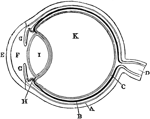
Diagram of the Eye | ClipArt ETC
Label the Eye Worksheet - Teacher-Made Learning Resources - Twinkl Here's a list of the main ones: Iris Sclera Pupil Lacrimal duct Cornea Lens Optic nerve Some of these are visible from the outside, like the iris and the pupil, but others would require a cross-section diagram to even see in the first place. Our Label the Eye worksheet covers all of these and more.

Structure Of Eye : External and Internal Structure of the Eyes
Eye Diagram Teaching Resources | Teachers Pay Teachers Use these simple eye diagrams to help students learn about the human eye. Three differentiated worksheets are included: 1. Write the words using a word bank2. Cut and paste the words3.

The Eyes (Human Anatomy): Diagram, Optic Nerve, Iris, Cornea ... - WebMD Articles On Eye Basics. Your eye is a slightly asymmetrical globe, about an inch in diameter. The front part (what you see in the mirror) includes: Iris: the colored part. Cornea: a clear dome ...

eye diagram
Human Eye Ball Anatomy & Physiology Diagram - eMedicineHealth The cornea is located just in front of the iris, which is the colored part of the eye. The main purpose of the cornea is to help focus light as it enters the eye. If one wears contact lenses, the contact lens rests on the cornea. Iris and Pupil. The iris, which is the colored part of the eye, controls the amount of light that enters the eye.

Label Parts of the Human Ear - University of Dayton Label Parts of the Human Ear. Select One Auditory Canal Cochlea Cochlear Nerve Eustachian Tube Incus Malleus Oval Window Pinna Round Window Semicircular Canals Stapes Tympanic Membrane Vestibular Nerve. Select One Auditory Canal Cochlea Cochlear Nerve Eustachian Tube Incus Malleus Oval Window Pinna Round Window Semicircular Canals Stapes ...

30 Label Eye - Labels For Your Ideas
Anatomy of the eye: Quizzes and diagrams - Kenhub Found within two cavities in the skull known as the orbits, the eyes are surrounded by several supporting structures including muscles, vessels, and nerves. There are 7 bones of the orbit, two groups of muscles (intrinsic ocular and extraocular), three layers to the eyeball … and that's just the beginning. There's a lot to learn, but stay calm!
Common Eye Problems and What They Look Like - LSC Eye Clinic
Anatomy of the Eye | Johns Hopkins Medicine Cornea. The clear, dome-shaped surface that covers the front of the eye. Iris. The colored part of the eye. The iris is partly responsible for regulating the amount of light permitted to enter the eye. Lens (also called crystalline lens). The transparent structure inside the eye that focuses light rays onto the retina. Lower eyelid.

34 Label The Parts Of Eye - Labels Database 2020
en.wikipedia.org › wiki › Human_eyeHuman eye - Wikipedia Each eye has seven extraocular muscles located in its orbit. Six of these muscles control the eye movements, the seventh controls the movement of the upper eyelid.The six muscles are four recti muscles – the lateral rectus, the medial rectus, the inferior rectus, and the superior rectus, and two oblique muscles the inferior oblique, and the superior oblique.

The Helpful Garden: Parts of the Eye Nomenclature Cards and Blackline Master Free Downloads ...
PDF Parts of the Eye Eye Diagram Handout Author: National Eye Health Education Program of the National Eye Institute, National Institutes of Health Subject: Handout illustrating parts of the eye Keywords: parts of the eye, eye diagram, vitreous gel, iris, cornea, pupil, lens, optic nerve, macula, retina Created Date: 12/16/2011 12:39:09 PM

Eye Anatomy
Eye Pictures, Anatomy & Diagram | Body Maps - Healthline Pads of fat and the surrounding bones of the skull protect them. The eye has several major components: the cornea, pupil, lens, iris, retina, and sclera. These work together to capture an image ...

Eye Facts for Kids
Shipping Labels, DOT Placards, UN Packaging from ... Jun 01, 2022 · Find all you need for hazmat shipping. Labelmaster offers UN packaging, CHEMTREC labels, GHS training, CFR's, DG shipping software, hazmat labels and placards and more.

› Sudden-Change-Under-Eye-FirmingAmazon.com: Sudden Change Under-Eye Firming Serum - Decreases ... Anti Ageing Eye Serum - Eye Cream - Anti Wrinkle Eye Serum for Puffy Eyes, Dark Circles, Eye Bags, Crows Feet, Wrinkles,Reduces Wrinkles Saggy Skin Puffy Eyes 4,083 $7.99 $ 7 . 99 ($7.99/Count) Sudden Change Green Tea Facial Mask – Diminish Wrinkles, Puffiness & More - Improve Texture, Purify Pores & Remove Excess Oil – Made with ...
Post a Comment for "39 parts of the eye without labels"